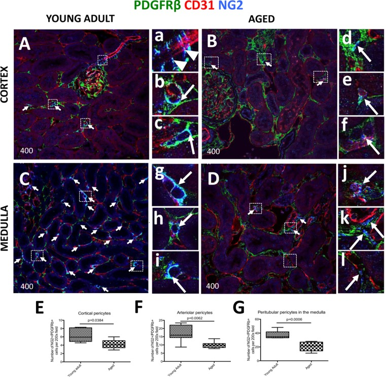
Figure 3.Decreased expression of kidney pericyte markers in aged micePericytes were identified by double NG2+/PDGFRß+ staining (blue and green colors respectively), and their perivascular location was identified by endothelial CD31 staining (red color). Double positive cells (light blue color) were quantified using single channel images (marked here by the arrows). (A) In young adult kidney cortex, staining for pericyte markers was readily detected alongside afferent arterioles (a, arrowheads) and peritubular capillaries (examples indicated in the boxed regions b and c, arrows). (B) In aged kidney cortex, the number of cells staining for pericyte markers and lining interstitial peritubular capillaries was reduced. Pericytes surrounding the vessels show loss of contacts with endothelial cells (d, arrow), and CD31 staining intensity was reduced (e, f, arrows). (C) Kidney medulla of young adult mice showed abundant pericytes located along peritubular capillaries. The boxed regions show higher power images of pericytes tightly encircling peritubular capillaries (g, h, i, arrows). (D) In aged kidney medulla, there was a decline in number of pericytes surrounding peritubular capillaries. Pericyte staining intensity diminished (examples are shown in the boxed regions j, k, l arrows), and the vessel coverage was lacking (k,l, arrows). In aged mice compared to young adults, pericyte number was decreased in: (E) peritubular capillaries in the cortex, (F) pre-capillary arterioles, (G) peritubular capillaries in the medulla. Data are represented as mean ± SEM (n=6).