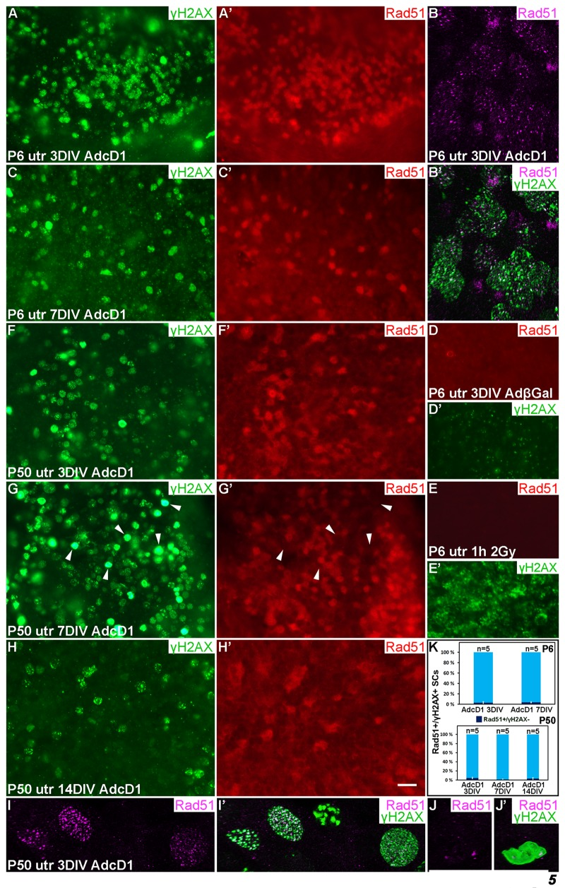
Figure 5.Dynamics of the DNA repair protein Rad51 in cell cycle reactivated supporting cells. Adenovirus-infected utricular explants were maintained for 3 to 14 DIV and double-labeled for γH2AX and Rad51. Imaging by conventional (A,A',C-H') and confocal microscopy (B,B',I-J'). (A,A') At 3 DIV, AdcD1-infected P6 utricle shows Rad51 upregulation in SCs with γH2AX foci. (B,B') High magnification view of an AdcD1-infected P6 utricle showing Rad51 expression as small foci that partially colocalize with the γH2AX foci. (C,C') By 7 DIV, the reduction in Rad51 levels is paralleled by γH2AX resolution. (D,D') AdβGal-infected P6 utricle lacks Rad51 expression. (E,E') P6 utricle exposed to ionizing radiation and analyzed 1 h post-irradiation contains SCs with DSB-like γH2AX foci. These cells lack Rad51 expression. (F,F') At 3 DIV, also the AdcD1-infected P50 utricle shows Rad51 induction in γH2AX+ SCs. (G,G') Rad51 expression is maintained at 7 DIV. Note that adult SCs with bright, pan-nuclear γH2AX staining lack or show only weak Rad51 expression (arrowheads). (H,H') Rad51 levels are reduced in the adult utricle by 14 DIV, paralleling γH2AX resolution. (I,I') High magnification view shows Rad51 foci in AdcD1-infected adult SCs and that these foci partially colocalize with γH2AX foci. (J,J´) High magnification view shows that adult SCs with pan-nuclear γH2AX staining express only low levels of Rad51. (K) Quantification shows that, at all timepoints studied and both in P6 and P50 utricles, Rad51 expression closely parallels that of γH2AX. Only 3-5% of Rad51+ SCs were negative for γH2AX. Mean ± SEM and the number of explants (n) are shown. Abbreviations: AdcD1, adenovirus encoding cyclin D1; AdβGal, adenovirus encoding β-galactosidase; γH2AX, Ser 139 phosphorylated histone H2AX; utr, utricle. Scale bar, shown in H': A,A',C-H', 20 µm; B,B',I-J', 5 µm.