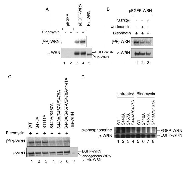
Figure 2.Bleomycin induces WRN phosphorylation at Ser-440 and 467 by DNA-PK(A-D) In vivo phosphorylation assay with radio-labeling. (A) Empty vector (pEGFP) (lanes 1 and 2) and pEGFP-WRN (lanes 3 and 4) were transfected to 293T cells. The cells were incubated in the absence (lanes 1 and 3) or presence (lanes 2 and 4) of 5 μg/ml bleomycin and [32P] labeled phosphate. WRN proteins were immunoprecipitated. Recombinant His-tagged full length WRN (800 ng) was loaded to assign the position of endogenous WRN (lane 5). The phosphorylated proteins were visualized (upper panel), followed by Western blotting with anti-WRN antibody (lower panel). (B) 293T cells transfected with pEGFP-WRN were treated with 5 μg/ml bleomycin in the presence of PI-3 kinase inhibitors, 25 μM wortmannin (lane 2) and 20 μM NU7026 (lane 3). (C) 293T cells transfected with pEGFP-WRN [wild type (WT) or mutant as indicated] were treated with or without 5 μg/ml bleomycin as indicated. Recombinant His-tagged full length WRN was loaded (lane 7). (D) In vivo phosphorylation assays using anti-phosphoserine antibody. HEK293 cells transfected with pEGFP-WRN (WT or mutant as indicated) were treated with (lanes 5-8) or without 5 μg/ml bleomycin (lanes 1-4),. EGFP proteins were immunoprecipitated with anti-GFP antibody. The phosphorylated proteins were detected by Western blotting with anti-phosphoserine antibody. The membrane was deprobed and analyzed by Western blotting with anti-WRN antibody.