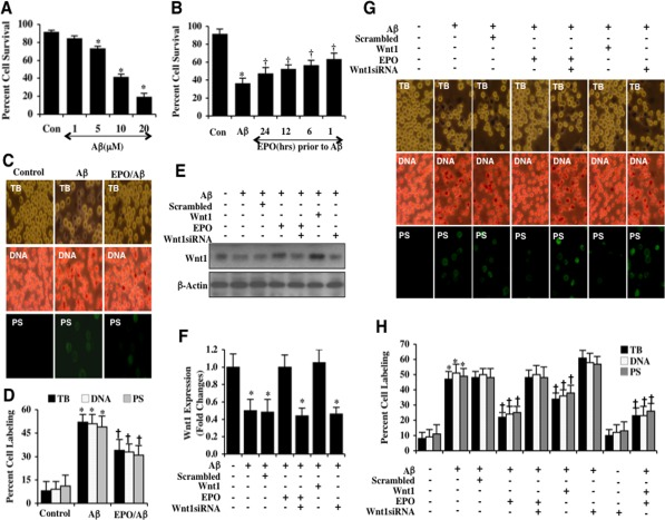
Figure 1.EPO preserves microglia survival against Aβ through Wnt1(A) Microglia were exposed to Aβ(10 μM) at the concentrations of 1, 5, 10, and 20 μM and cell survival was determined 24 hours after administration of Aβwith trypan blue dye exclusion method (*P< 0.01 vs. Control). Con = control = untreated microglia. Each data point represents the mean and SEM from 6 experiments. (B) EPO (10 ng/ml) was applied to microglial cultures at 24, 12, 6, or 1 hour prior to administration of Aβ (10 μM) and cell survival was determined 24 hours after Aβ administration with the trypan blue dye exclusion method (*P<0.01 vs. untreated control; †P <0.05 vs. Aβ). Each data point represents the mean and SEM from 6 experiments. Con = control = untreated microglia. (C and D) EPO (10 ng/ml) was applied to microglial cultures 1 hour prior to the administration of Aβ (10 μM) and cell survival, DNA fragmentation, and PS exposure were determined 24 hours later. Representative images (C) and quantitative analysis (D) demonstrate that Aβ leads to a significant increase in trypan blue staining, DNA fragmentation, and membrane PS exposure in microglia 24 hours after Aβ exposure compared to untreated control cultures. EPO (10 ng/ml) applied 1 hour prior to Aβ exposure prevented microglial cell injury, DNA fragmentation, and membrane PS exposure(*P < 0.01 vs. Control; †P <0.05 vs. Aβ). Each data point represents the mean and SEM from 6 experiments. (E and F) EPO (10 ng/ml) or Wnt1 (100 ng/ml) were applied 1 hour prior to Aβ (10 μM) administration with Wnt1 expression determined 6 hours following Aβ exposure. EPO (10 ng/ml) and Wnt1 (100 ng/ml) maintained the expression of Wnt1 that is otherwise down-regulated during Aβ exposure. Gene reduction ofWnt1with Wnt1 siRNA significantly reduced the expression of Wnt1 following a 6 hour period of Aβ exposure or treatment with EPO (10 ng/ml) during Aβ exposure. Non-specific scrambled siRNA did not alter Wnt1 expression during Aβ exposure (*P<0.01 vs. Control). (G and H) EPO was applied to microglial cultures 1 hour prior to the administration of Aβ and trypan blue dye exclusion, DNA fragmentation, and membrane PS exposure were determined 24 hours later. Representative images (G) and quantitative results (H) show that EPO (10 ng/ml) or Wnt1 (100 ng/ml) applied 1 hour prior to Aβ significantly reducedtrypan blue staining, DNA fragmentation, and membrane PS exposure in microglia 24 hours after Aβ exposure. Gene reduction of Wnt1 with transfection of Wnt1 siRNA prior to Aβ exposure prevented EPO (10 ng/ml) from blocking cell injury and resulted in increased trypan blue staining, DNA fragmentation, and membrane PS exposure in microglia 24 hours following Aβ exposure. Non-specific scrambled siRNA did not significantly alter microglial cell injury following Aβ exposure (*P<0.01 vs. untreated control; †P < 0.05 vs. Aβ). Each data point represents the mean and SEM from 6 experiments.