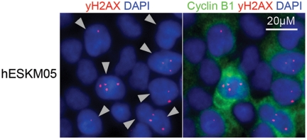
Figure 4.Double immunofluorescence staining of hESCs with γ-H2AX (red) and G2-specific Cyclin B1 (green) antibodies revealed high frequency of γ-H2AX foci in cells at G2 phase.Nuclei were counterstained with DAPI (blue). Gray triangles indicate G2-cells.