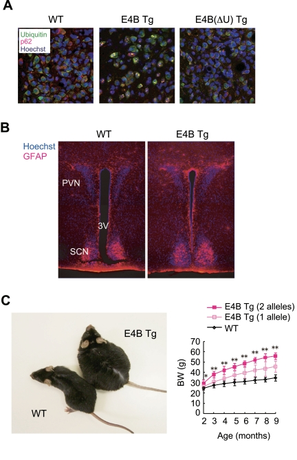
Figure 1. E4B
transgenic (Tg) mice as a new obesity model with hypothalamic
neurodegeneration. (A) Immunofluorescence analysis
of the PVN region of 6-month-old wild-type (WT) or E4B(ΔU) Tg mice and
of a 4-month-old E4B Tg mouse. Brain slices were stained with antibodies to
polyubiquitin (green) and to p62 (red), and nuclei were stained with
Hoechst 33258 (blue). Protein aggregates reacted with both types of
antibody in the PVN region of E4B Tg mice, but not in that of WT or
E4B(ΔU) Tg mice. (B) Immunofluorescence analysis of the PVN
region of 10-week-old WT or E4B Tg mice with antibodies to glial fibrillary
acidic protein (GFAP, red). Nuclei were stained with Hoechst 33258 (blue).
SCN and 3V indicate the suprachiasmatic nucleus and third ventricle,
respectively. The number of GFAP-positive glial cells in and around the PVN
was increased in E4B Tg mice, indicative of gliosis associated with
neurodegeneration. (C) Obesity in E4B Tg mice. The gross appearance
of an E4B Tg mouse and a WT littermate at 9 months of age is shown on the
left. The time course of body weight (BW) for WT mice and E4B Tg lines
harboring one or two alleles of the transgene is shown on the right. The
extent of obesity in the Tg animals harboring two alleles of the transgene
was about twice that in littermates harboring only one allele, indicating
that the obese phenotype is directly related to the expression level of the
transgene. *P < 0.05, **P < 0.01 for the Tg line with
two alleles of the transgene versus wild-type mice.