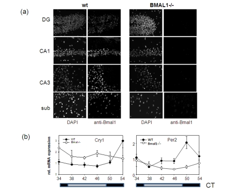
Figure 2.Expression of circadian proteins in brain structures.(a)
Immunostaining of sagittal brain sections of wt and Bmal-/- mice
with BMAL1 specific antibodies. Counterstaining with DAPI was used to
detect nuclei. Pyramidal neurons of hippocampal areas CA1 and CA3, granular
cells of the dentate gyrus and neurons of subiculum expressing BMAL1 are
shown. (b) Circadian profile of Cry1 and Per2 mRNAs in
the brain of wt (filled circles) and Bmal1-/- mice (open circles) as
measured by real-time PCR. (* p<0.05).