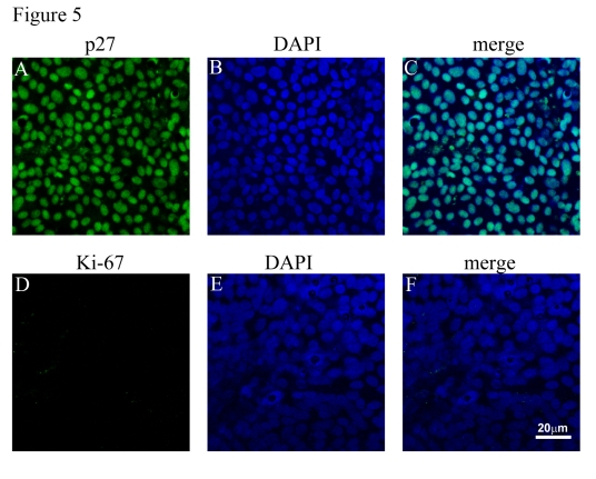
Figure 5.Cell cycle analysis of polarized RPE monolayers. (A, B, C)
Expression of p27 (green) and its localization to nuclei (blue). (D, E,
F). Polarized RPE cultures show lack of expression of Ki-67 (green) in
the nuclei. Nuclei counterstained blue with DAPI.