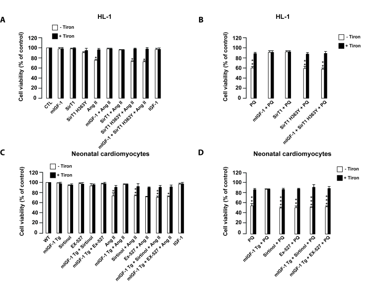
Figure 8.mIGF-1 prevents Ang II- and PQ-dependent cell death in HL-1 cardiomyocytes and in mouse neonatal primary cardiomyocytes.(A, B)
HL-1 cardiomyocytes were transfected or treated as in Legend of Figure 2A. (C, D)
Neonatal primary cardiomyocytes from wild type or mIGF-1 Tg mice were
treated as in Legend of Figure 2B. (A-D) Cell viability was
monitored with propidium iodide (PI) by flow cytometry and values were
normalized to protein content. Results are means ± SE of 3 independent experiments
(*,**,***p versus unstimulated
control cells or untreated wild type cardiomyocytes).