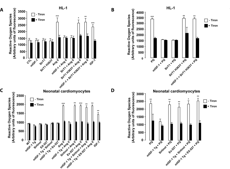
Figure 6.mIGF-1 prevents Ang II-, PQ- and IGF-1-induced increase in reactive oxygen species (ROS) generation in HL-1 cardiomyocytes and in mouse neonatal primary cardiomyocytes.(A,
B) HL-1 cardiomyocytes were transfected or treated as in Legend of
Figure 2A, except that Ang II (1 μM)
or PQ (100 μM) were added
for only 60 min. Untransfected cells were used as control (CTL). (C, D) Neonatal
primary cardiomyocytes from wild type or mIGF-1 Tg mice were treated as in
Legend of Figure 2A, except that Ang II (1 μM) or PQ (100 μM)
were added for only 60 min. (A-D) ROS production was monitored with
the fluorescent probe dichlorofluorescein diacetate (CM-DCFDA) and
fluorescence values were normalized to protein content. Results are means ± SE of 3 independent experiments
(*,**,***p versus unstimulated
control cells or untreated WT cardiomyocytes).
Figure 6 — Local IGF-1 isoform protects cardiomyocytes from hypertrophic and oxidative stresses via SirT1 activity | Aging