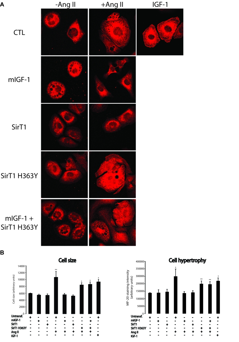
Figure 3.mIGF-1 prevents Ang II- and IGF-1-induced cell hypertrophy (MF-20 staining) in HL-1 cardiomyocytes.(A) HL-1 cardiomyocytes were
transfected or treated as in Legend of Figure 2A. Sarcomeric myosin was
stained with MF-20 antibody and images were acquired using a Leica confocal
microscope. (B) Cell size and cell hypertrophy quantified according
to MF-20 staining in HL-1 cardiomyocytes in the different experimental
conditions as in as in Legend of Figure 2A. Results are means ± SE of 3 independent experiments
(*,**p versus unstimulated
control cells). Bar: 25 μM.