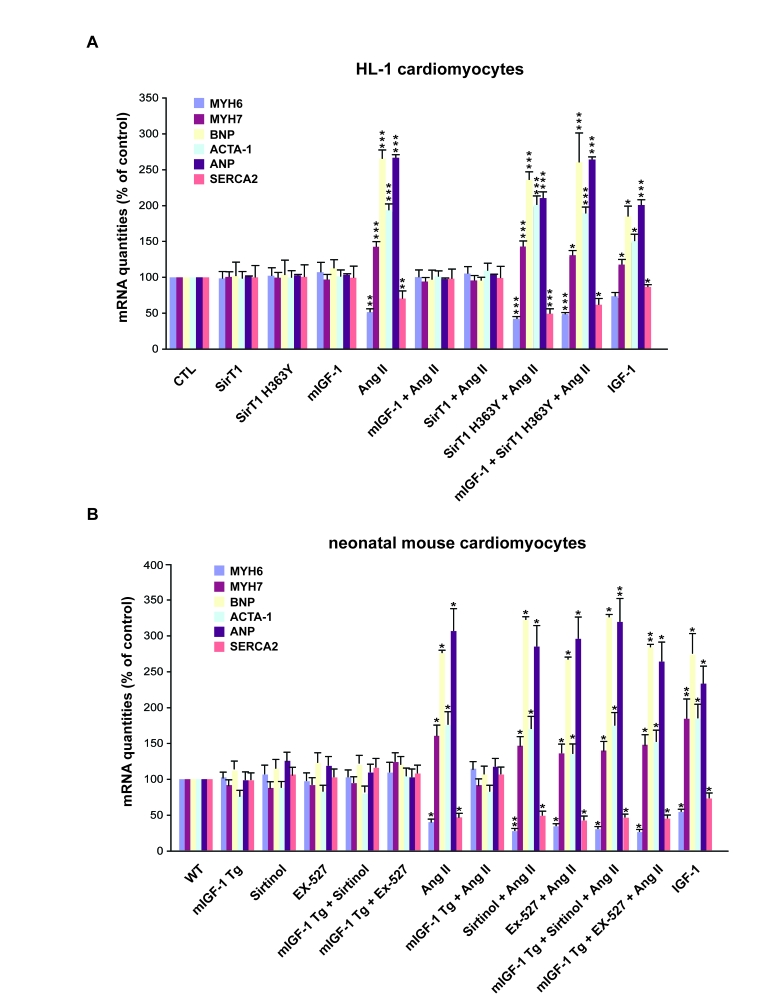
Figure 2.mIGF-1 prevents Ang II- and IGF-1-induced fetal gene program activation.(A) HL-1
cardiomyocytes were transfected with the indicated plasmids, or treated
with 20 ng/ml IGF-1 for 24 h, before exposure to Ang II (1 μM for 24 h).
Untransfected cells were used as control (CTL). (B) Neonatal mouse
cardiomyocytes from wild type (WT) or heart overexpressing mIGF-1 mice
(mIGF-1 Tg) were pre-incubated with sirtinol (100 μM) or EX-527 (1 μM), or
treated with 20 ng/ml IGF-1 for 24 h, prior to exposure to Ang II (1 μM for
24 h). Untreated WT cardiomyocytes were used as control. (A, B) The
expression levels of MYH6, MYH7, BNP, ACTA-1, ANP and SERCA2 mRNAs were
examined by qRT-PCR. Results are means ±
SE of 3 independent experiments (*,**,***p versus unstimulated
control cells).