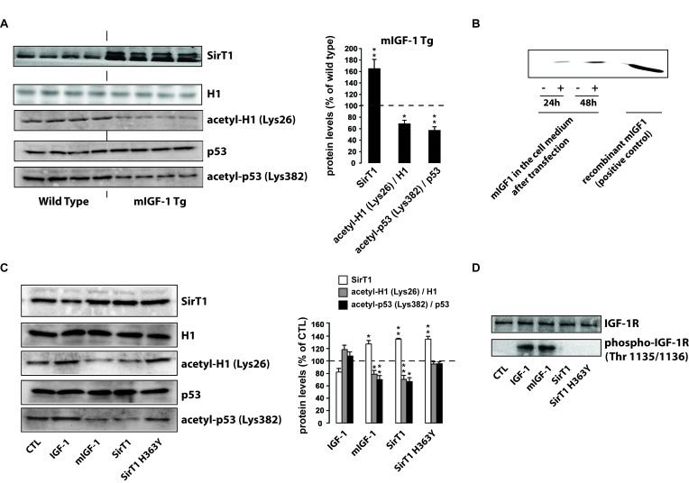
Figure 1.mIGF-1, but not IGF-1, increases SirT1 expression and activity in mouse cardiomyocytes. (A) Left
panel: representative Western blots of SirT1, histone H1, acetyl-H1
(Lys26), detected in nuclear extracts, and of p53 and acetyl-p53 (Lys382),
detected in whole tissue lysates, from wild type and mIGF-1 Tg mice. Four
animals of a total of 10 are shown; right panel: densitometric
quantification of SirT1, acetyl-H1(Lys26)/H1 and acetyl-p53(Lys382)/p53
levels in cardiomyocytes from mIGF-1 mice, expressed as % of those in wild
type cardiomyocytes. (B) representative Western Blot of mIGF-1
detected in the extracellular medium of HL-1 cardiomyocytes, transfected
with a plasmid carrying mouse mIGF-1 cDNA. (C) Left panel:
representative Western Blot of SirT1, histone H1, acetyl-H1 (Lys26)
detected in nuclear extracts, and of p53 and acetyl-p53 (Lys382) detected
in whole cell lysates, from HL-1 cardiomyocytes transfected with the
indicated constructs (SirT1 or SirT1 H363Y) or treated with 20 ng/ml IGF-1
for 24 hours; right panel: densitometric quantification of SirT1,
acetyl-H1(Lys26)/H1 and acetyl-p53(Lys382)/p53 levels in transfected or
treated cells, expressed as % of control (CTL). (D) Representative
Western blots of IGF-1 receptor (IGF-1R) or phospho-IGF-1R (on Thr
1135/1136) in HL-1 cardiomyocytes lysates. Results in (A) and (B)
are means ± SE of 3
independent experiments (**,***p versus unstimulated
control cells or untreated WT cardiomyocytes).