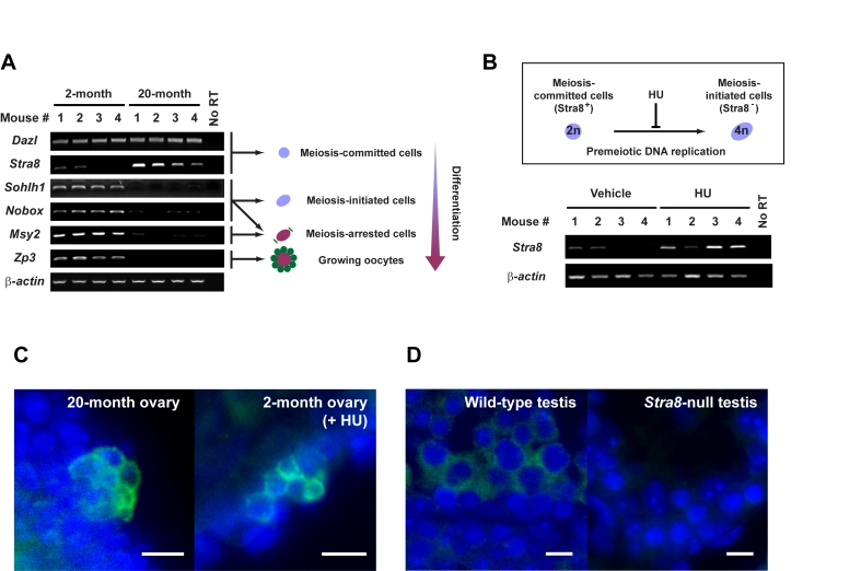
Figure 1.Premeiotic germ cells persist in aged atrophic mouse ovaries.
(A) Analysis of germline marker gene expression in ovaries of young adult
(2-month) and aged (20-month) female mice. Results from all 4 mice per age
group are shown (β-actin, housekeeping
gene used as a sample loading control). (B) In-vivo blockade of
premeiotic DNA replication by HU in ovaries of young adult mice results in
enhanced levels of Stra8 expression, consistent with premeiotic germ
cell accumulation. (C) Immunofluorescence analysis of STRA8
expression (green, cytoplasm) in ovaries of aged or HU-treated young
adult female mice. (D) Control immunofluorescence analysis of STRA8
expression (green, cytoplasm) in testes of young adult wild-type or Stra8-null
male mice (a representative cross-section of seminiferous tubule is shown
for each.). C, D: scale bar = 10 μm;
DAPI counterstain, blue (nucleus).