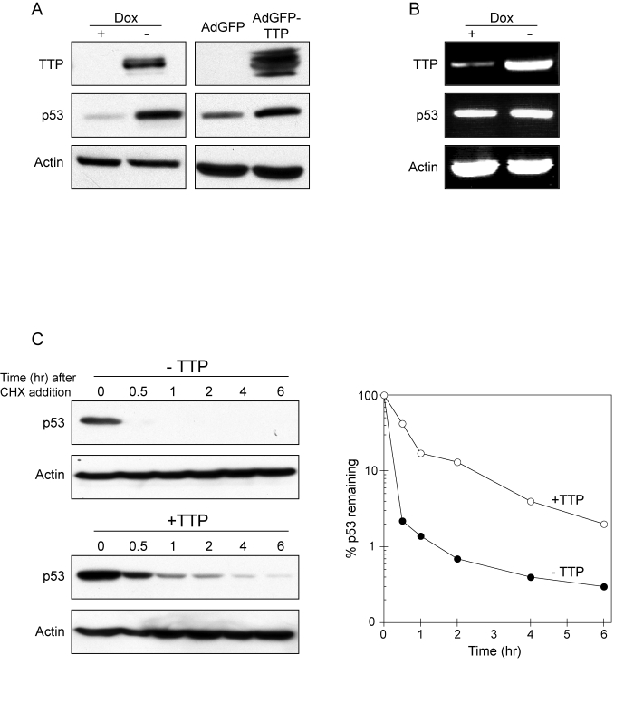
Figure 2.TTP promotes p53 expression through protein stabilization.
(A) HeLa Tet-Off/TTP-Flag cells grown in presence or absence of Dox for 48 hr
(left panel) and HeLa cells infected with AdGFP or AdGFP/TTP virus for 48 hr
(right panel) were examined for TTP and p53 expression by western blotting.
Actin was used as a loading control. (B) RT-PCR analysis of p53 mRNA
expression in HeLa Tet-Off/TTP-Flag cells grown in presence or absence of
Dox for 48 hr. Induction TTP-Flag mRNA is shown along with loading control
GAPDH. (C) TTP promotes
increased stability of p53 protein. HeLa Tet-Off/TTP-Flag cells grown in
presence (- TTP) or absence (+ TTP) of Dox for 48 hr were incubated with 20
μg/ml cycloheximide (CHX) to inhibit
protein synthesis for the indicated times. Decay of p53 protein was
examined by western blot (left panels) using actin as a loading control.
Decay curves of p53 protein (right panel) in the presence (open circles)
and absence (filled circles) of TTP was obtained by western blot analysis
and normalized to the internal control actin.