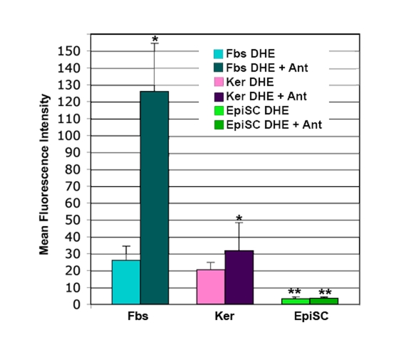
Figure 3.DHE staining of superoxide in skin cells.Cultures of
dermal fibroblasts (Fbs), epidermal keratinocytes (Ker), and epidermal stem
cells (EpiSC) were stained with dihydroethidium (DHE) in the presence or
absence of the electron transport chain blocker antimycin A (Ant).
Fluorescence for each cell type +/- Ant was determined by flow cytometry,
then normalized by comparison to a standard cell. EpiSCs show significantly
lower levels of DHE staining than the keratinocyte and fibroblast
populations (p<0.01). Increase in DHE staining in the Fbs+Ant samples
was significantly higher than that seen in Ker+Ant samples (p<0.05).
Lack of increase in DHE staining in the EpiSC after antimycin A treatment
was significant (p<0.01). Significant differences were determined by student's
T-test. n=5.