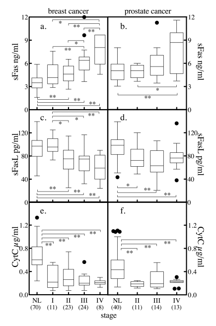
Figure 6.Serum markers of apoptosis and tumor stage. Subjects with
breast cancer (a, c, e), or prostate cancer (b, d, f) were
stratified by stage and the distribution of sFas (a, b), sFasL (c,
d) and cytochrome c (e, f)
stratified by staging was determined. The solid horzontal bars depict the
median values. For breast cancer, stage I tumor size (T) < 2 cm across
and cancer cells have not spread to axillary lymph nodes (N). For stage II,
T < 2 cm across and the cancer has spread to the lymph nodes under the
arm (N positive) or T is 2 to 5 cm and N is negative. In stage III, T >
5 cm or it has spread to other lymph nodes or tissues near the breast.
Stage IV is metastatic cancer. The convention for prostate cancer staging
was that in stage I, cancer is found in the prostate only. In stage II,
cancer is more advanced than in stage I, but has not spread outside the
prostate. In stage III, cancer has spread beyond the outer layer of the
prostate to nearby tissues. Stage IV is characterized by distant
metastasis. Comparison between group median values was performed by Mann
Whitney t-test, where * = p < 0.05, ** = p < 0.005, *** = p <
0.0001. Numbers in parenthesis indicate number of subjects in each group.