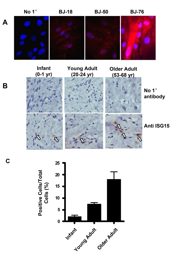
Figure 5.ISG15 is increased in human skin with aging. (A)
Immunofluorescence staining of ISG15 in BJ cells at different population
doublings. The negative sample was treated identically except no primary
antibody was added. Nuclei were stained with DAPI. Staining intensity
increases in cells with short telomeres. (B) Immunochemical staining
illustrating an age-dependent up-regulation of ISG15 expression in the
dermis of human skin tissues. 2-4 cases were examined in each group.
Infant, 0-1 year old; young adult, 20-24 year old; older adult, 53-68 year
old. No primary antibody was added to the negative control. (C)
Quantitation of the results of all the samples described above. 8-10 random
fields were counted for each sample.