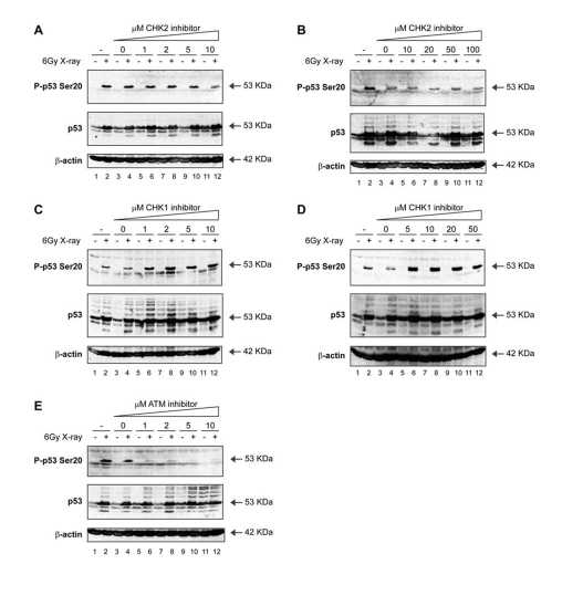
Figure 4.Activation of p53 by ionising radiation: effects of ATM-CHK pathway inhibitors on p53 phosphorylation. (A, B)
A CHK2 inhibitor does not attenuate Ser20 site phosphorylation of
p53 nor p53 induction mediated by treatment with
X-rays. MOLT-3 cells were treated with (even-numbered lanes)
or without (odd-numbered lanes) 6Gy X-ray and cultured for 4
hours after an initial 44-hour pre-treatment with: increasing
concentrations [1-10μM (A) or 10-100μM (B)] of the CHK2 inhibitor
(lanes 5-12), a DMSO solvent control (lanes 3-4), or a culture
medium control (lanes 1-2). Cell lysates were examined by Western
blotting with antibodies against the indicated proteins.
(C, D) A CHK1 inhibitor does not attenuate Ser20 site phosphorylation
of p53 nor p53 induction mediated by treatment with X-rays.
MOLT-3 cells were treated with (even-numbered lanes) or without
(odd-numbered lanes) 6Gy X-ray and cultured for 4 hours after
an initial 44-hour pre-treatment with: increasing concentrations
[1-10μM (C) or 5-50μM (D)] of the CHK1 inhibitor SB218078
(lanes 5-12), a DMSO solvent control (lanes 3-4), or a culture
medium control (lanes 1-2). Cell lysates were examined by Western
blotting with antibodies against the indicated proteins.
(E) An ATM inhibitor attenuates Ser20 site phosphorylation of p53,
but not p53 induction, mediated by treatment with X-rays. MOLT-3
cells were treated with (even-numbered lanes) or without
(odd-numbered lanes) 6Gy X-ray and cultured for 4 hours after
an initial 44-hour pre-treatment with: increasing concentrations
(1-10μM) of the ATM inhibitor KU-55933 (lanes 5-12), a DMSO
solvent control (lanes 3-4), or a culture medium control
(lanes 1-2). Cell lysates were examined by Western blotting
with antibodies against the indicated proteins.