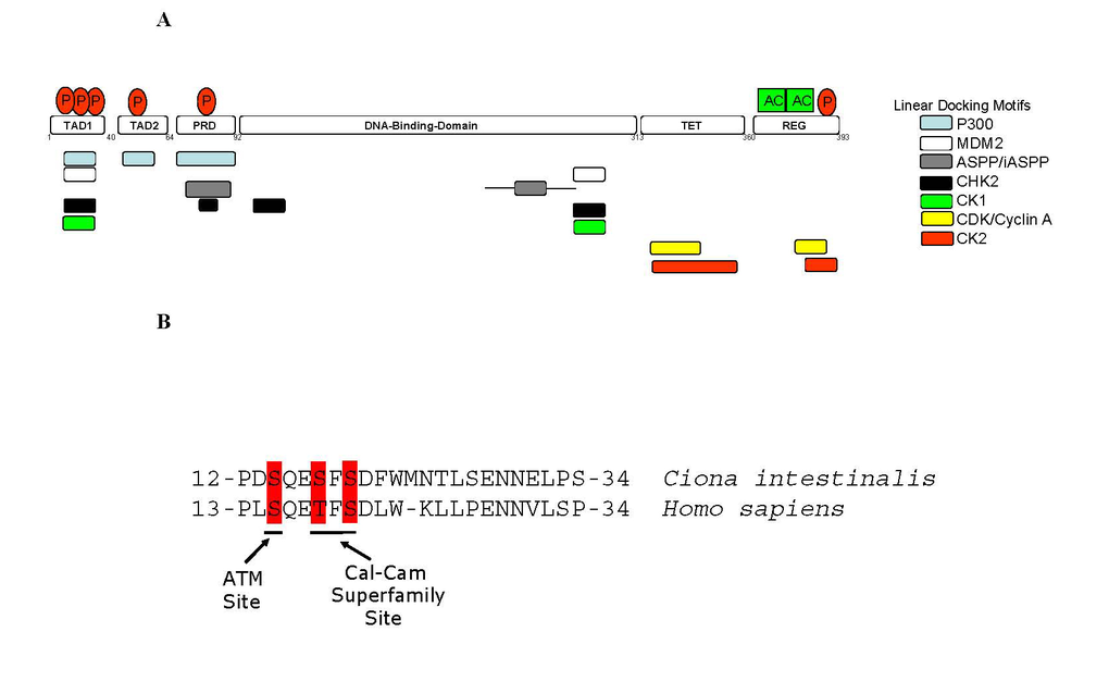
Figure 2.Linear Peptide Docking Sites in p53.
(A) Linear peptide docking
sites for enzymes that regulate p53 function. The N-terminus
is composed of three transactivation motifs,TAD1, TAD2, and Proline-repeat
domain (PRD). A key regulatory domain in the C-terminus (REG) contains the
acetylation motifs and phosphorylation site and flanks the Tetramerization
domain (TET). The overlapping, but distinct, linear polypeptide docking
motifs for p53 regulators include the acetyltransferase p300, the E3
ubiquitin ligase MDM2, iASPP, and the protein kinases including CDK, CK2,
CK1, and CHK2 are highlighted. (B) Conservation of key
phospho-acceptor sites between urochordate and human. The panel
highlights the conservation of amino acids and phospho-acceptor sites in
the BOX-I transactivation domain of p53 (TAD1 in Figure 2A) between human and urochordate
(Ciona intestinalis).
The ATM phospho-acceptor site at Ser15 and the Calcium Calmodulin kinase/CK1
phospho-acceptors sites at Thr18 and Ser20 are highlighted as indicated.