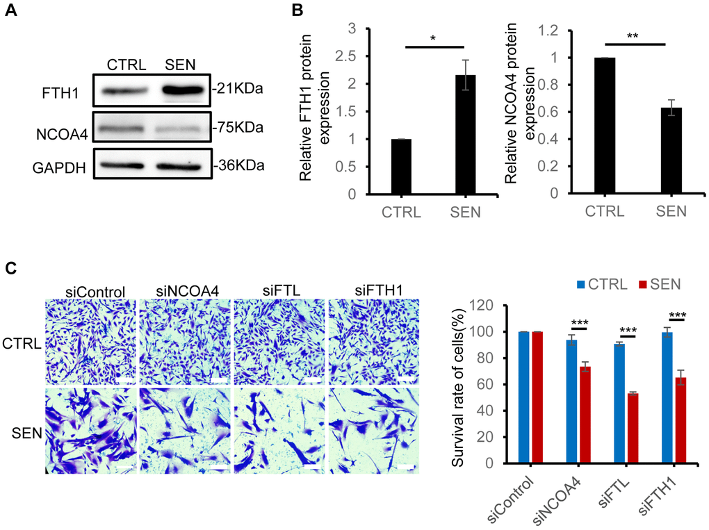
Figure 5.Disruption of ferritinophagic process promotes senescent cell death. (A) Immunoblot analysis of FTH1, NCOA4 protein expression in proliferation and senescent HSkM cells. (B) Quantification of the ratio of FTH1 and NCOA4 protein expression. Each protein band intensity was measured and normalized to GAPDH, compared between proliferating and senescent cells. Data were collected from three independent biological replicates, and represented as mean ± SD. P-values were calculated by two-tailed unpaired student’s t-test, *P < 0.05. (C) FTH1, FTL and NCOA4 knockdown decreased the survival rate of senescent cells. The representative field images (left) and quantification of cell survival ratio (right). Cells were seeded on 24-well plate and the respective genes were knockdown by their specific siRNA for 48 h, followed by crystal violet staining. The cells were viewed by an inverted light microscope and counted the number of cells in different fields. The plate image is presented in Supplementary Figure 4E. Data were collected from three independent biological replicates, and represented as mean ± SD. P-values were calculated by one-way ANOVA analysis with post hoc Tukey, ***P < 0.001.