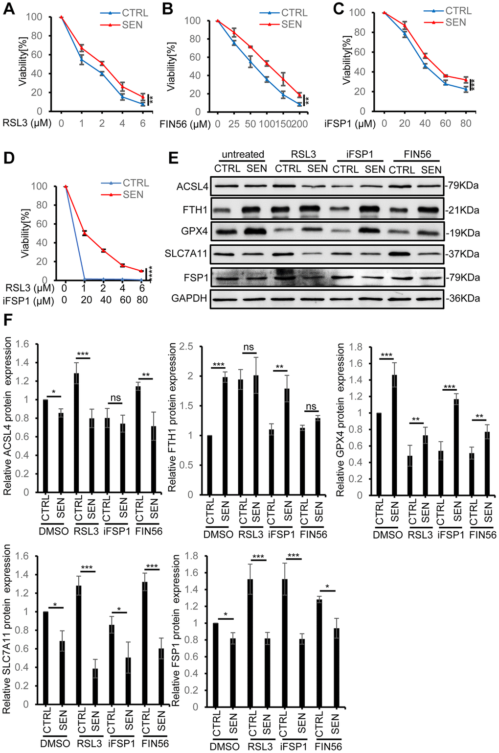
Figure 3.Senescent HSkM cells are resistant to ferroptosis inducers. (A–C) Senescent HSkM cells are more resistant to ferroptosis inducers RSL3, FSP1 and FIN56. The proliferating and senescent HSkM cells were treated with a series of concentrations of RSL3, FSP1 and FIN56 for 24 h, respectively. (D) Senescent cells are more resistant to the synergistical treatment of RSL3 and iFSP1. Cell viability was determined by CCK-8 assay. P-values were calculated by two-way ANOVA analysis, **P < 0.01, ***P < 0.001. Data represented as mean ± SD. And three independent biological experiment repeats have been performed. (E, F) The expression of ferroptosis-related proteins in proliferating and senescent HSkM cells under RSL3, FIN56 and iFSP1 treatment. Immunoblot representative images (E) and quantification (F) of ACSL4, FTH1, GPX4, SLC7A11, FSP1 protein expression in normal and senescent HSkM cells after treatment with RSL3, iFSP1, FIN56, respectively. Each protein band intensity was measured and normalized to GAPDH and compared between proliferating and senescent cells. Data represented as mean ± SD. And three independent biological experiment repeats have been performed. P-values were calculated by one-way ANOVA analysis with post hoc Tukey, *P < 0.05, **P < 0.01, ***P < 0.001.