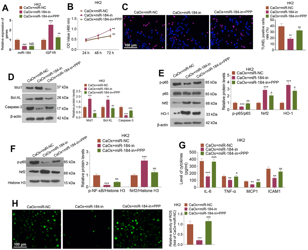
Figure 7.IGF1R pathway inhibition reversed the inhibitory impact of miR-184 knockdown on renal cell impairment. Following transfection with miR-184-in and intervention with IGF1R pathway inhibitors (PPP, 1 μMnM), HK2 cells were treated with CaOx (100 μg/mL) for 24 hours. (A) The profiles of miR-184 and IGF1R were determined by qRT-PCR. (B) A CCK8 assay was performed to examine cell viability. (C) Cell apoptosis was monitored through TUNEL staining. Scale bar=100 μm. (D) Western blot analysis of the profiles of apoptosis-associated proteins. (E, F) Western blot analysis verified the profiles of the NF-κB and Nrf2/HO-1 pathways. (G) The levels of the inflammatory cytokines IL-6, TNF-α, MCP1, and ICAM1 were determined by ELISA. (H) Immunofluorescence evaluated the activity of ROS. Scale bar=100 μm. *P<0.05, **P<0.01, ***P<0.001 (vs. CaOx+miR-in). nsP>0.05, ^P<0.05, ^^P<0.01 (vs. CaOx+miR-184-in). N=3.