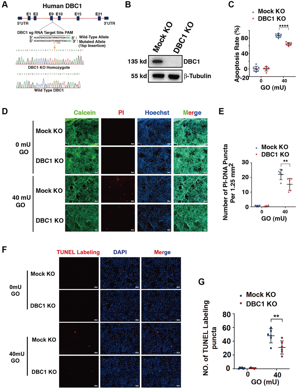
Figure 5.DBC1 knockout significantly attenuates glucose oxidase (GO)-induced apoptosis. (A) A schematic diagram showing strategy for DBC1 knockout in FHL124 cells by CRISPR/Cas9 gene editing technology. The red underlined base pairs are the sgRNA target, the green underlined base pairs are the protospacer-adjacent motif (PAM). 1-bp base insertion in the mutated allele are shown in red bold. The stop codon introduced in the mutant form is shown. (B) Western blot analysis of DBC1 expression levels in control (Mock KO) and DBC1 knockout (DBC1 KO) cells. Note that expression of DBC1 was not detectable in DBC1 knockout cells. The β-tubulin served as the loading control. (C) Apoptosis rate changes in Mock KO and DBC1 KO cells under treatment of 40 mU GO for 5 hours were measured by CellTiter-Lumi™ II Luminescent Cell Viability assay analysis. (D) Calcein/PI Cell Viability/Cytotoxicity assay analyzed cell apoptosis of Mock KO and DBC1 KO cells under the same treatment as in C. Green fluorescence represents live cells as detected by Calcein-AM, and red fluorescence detected by PI refers to dead cells. Scale bar, 100 μm. (E) Quantification of the PI-DNA puncta in panel D. **p < 0.01, ****p < 0.0001. (F) TUNEL labeling assay under the same treatment as in C. Scale bar, 100 μm. (G) Quantification of the TUNEL Labeling puncta in panel F. **p < 0.01.