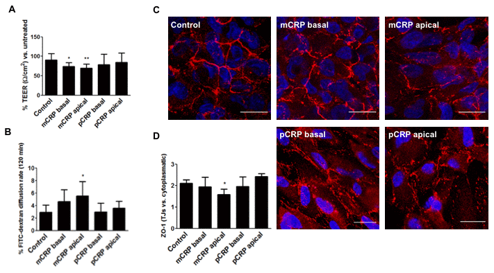
Figure 5.mCRP induces barrier disruption in ARPE-19 cells in a polarized manner. ARPE-19 cells were treated with CRP isoforms for 48h either from the apical side or the basolateral chamber and TEER (A) and paracellular permeability as determined by FITC-dextran diffusion rate (B) was determined. (C) Cells were then fixed and immunostained with anti ZO-1 (red) and DAPI (blue). Images shown are representative of four independent experiments. Scale bar = 20 μm. (D) Quantification of ZO-1 at the TJs expressed as relative (intercellular/cytoplasmic) ZO-1 distribution. Values are expressed as mean ± SD and statistical analysis was performed by one-way ANOVA and Dunnett´s posthoc analysis (N=4). * P<0.05, ** P<0.01 vs. control.